Health
Claude Horner: The History, Medical Significance, and Lasting Legacy

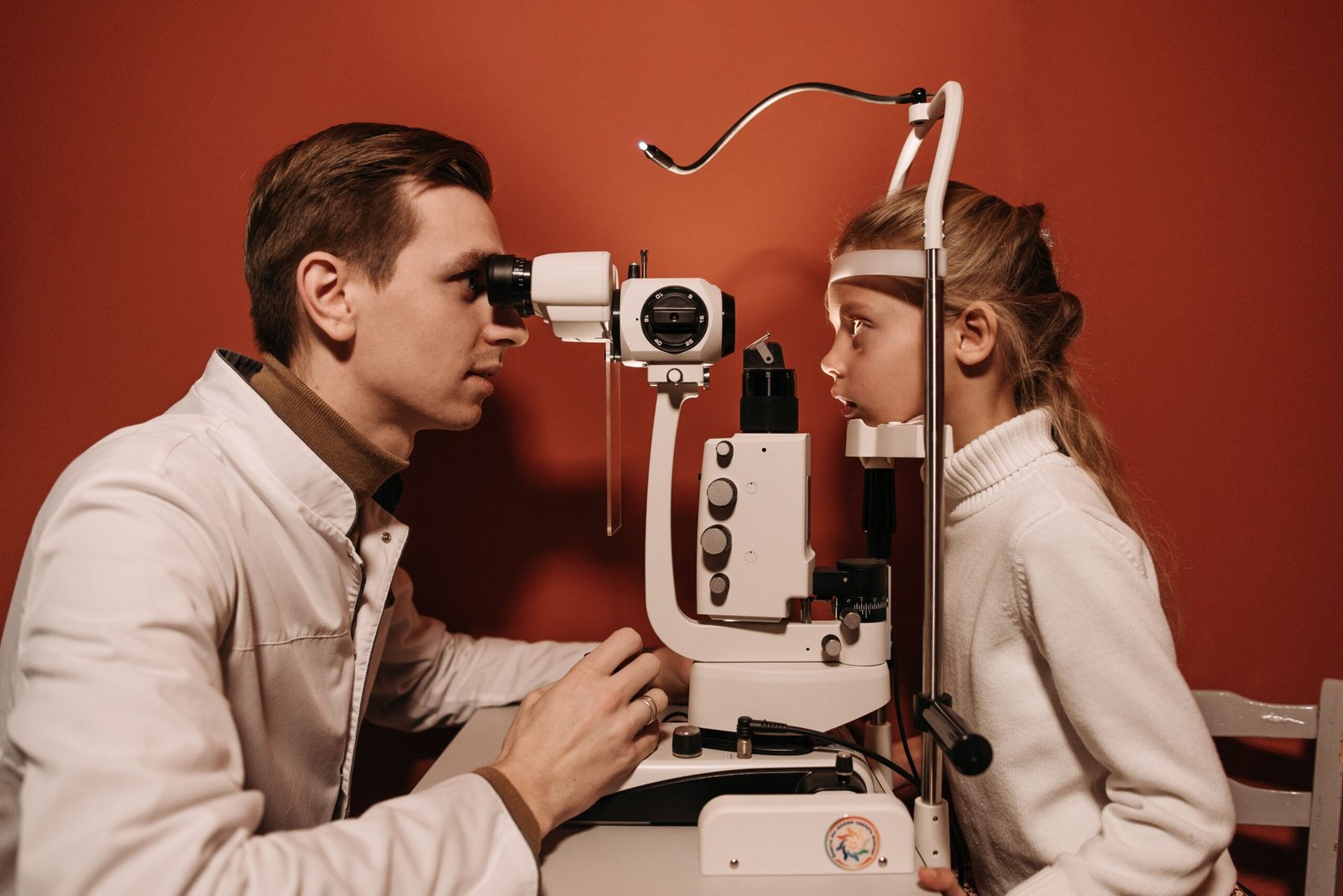
The term Claude Horner is often associated with one of the most important neurological and ophthalmological conditions in medical history: the Claude Bernard-Horner syndrome. Although the name combines contributions from multiple medical pioneers, it represents a fascinating story of scientific discovery, clinical observation, and advances in understanding the human nervous system.
For more than a century, physicians, neurologists, ophthalmologists, and medical researchers have studied the condition known as Horner syndrome. The syndrome provides valuable insight into the sympathetic nervous system and remains an important diagnostic clue for various neurological disorders.
Understanding the history behind Claude Horner, the individuals connected to the syndrome, and its impact on modern medicine helps illustrate how scientific observations can influence healthcare for generations. This article explores the origins, discoveries, clinical significance, symptoms, diagnosis, treatment, and ongoing relevance of the Claude Bernard-Horner syndrome.
Quick Facts
| Attribute | Details |
|---|---|
| Medical Name | Claude Bernard-Horner Syndrome |
| Common Name | Horner Syndrome |
| Condition Type | Neurological Disorder |
| Named After | Claude Bernard and Johann Friedrich Horner |
| First Described | 19th Century |
| Affected Area | Eye, Face, Neck |
| Main Cause | Damage to Sympathetic Nerves |
| Common Symptoms | Miosis, Ptosis, Anhidrosis |
| Eye Involvement | Usually One Eye |
| Facial Involvement | Usually One Side |
| Vision Loss | Rare |
| Pain Association | Possible in Some Cases |
| Diagnosis Method | Clinical Examination and Imaging |
| Treatment Focus | Treating Underlying Cause |
| Emergency Cases | Possible with Stroke or Artery Dissection |
| Age Group | Any Age |
| Congenital Cases | Yes |
| Adult Cases | Common |
| Recovery Potential | Depends on Cause |
| Medical Specialty | Neurology and Ophthalmology |
The Origins of the Claude Horner Name
The phrase Claude Horner is closely linked to the historical development of Horner syndrome. The syndrome is commonly known as Claude Horner syndrome in parts of Europe because it honors both the French physiologist Claude Bernard and the Swiss ophthalmologist Johann Friedrich Horner.
Claude Bernard conducted groundbreaking experiments involving the sympathetic nervous system during the mid-19th century. His research demonstrated how interruption of sympathetic nerve pathways affected the eye and facial structures. Later, Johann Friedrich Horner provided a detailed clinical description of the syndrome in 1869, helping establish the condition’s diagnostic significance.
Because of these combined contributions, the condition became known in many medical circles as Claude Bernard-Horner syndrome, leading to the modern association of the term Claude Horner.
Understanding Horner Syndrome and Its Medical Importance
Horner syndrome is a neurological condition caused by disruption of the sympathetic nerve pathway that connects the brain to the face and eye. The syndrome typically presents with a distinct set of symptoms that affect one side of the face.
The sympathetic nervous system controls several automatic bodily functions, including pupil dilation, eyelid position, sweating, and blood vessel regulation. When this pathway becomes damaged, characteristic signs appear that often help physicians identify underlying medical conditions.
The syndrome remains clinically important because it is frequently a symptom rather than a disease itself. In many cases, Horner syndrome serves as a warning sign that prompts further investigation into potentially serious neurological, vascular, or structural disorders.
The continued relevance of Claude Horner in medical education demonstrates the enduring value of historical scientific discoveries.
The Scientific Contributions of Claude Bernard and Johann Friedrich Horner
One of the most fascinating aspects of Claude Horner is the collaboration of scientific ideas across different generations.
Claude Bernard’s Experimental Discoveries
Claude Bernard was among the most influential physiologists in medical history. During the 1850s, he conducted experiments that revealed the role of the cervical sympathetic nerves in controlling eye function and facial responses.
His observations showed that damage to sympathetic nerve pathways produced noticeable changes in the eye, including pupil constriction and eyelid abnormalities. These findings helped establish a foundation for modern neurophysiology.
Johann Friedrich Horner’s Clinical Description
Johann Friedrich Horner expanded upon Bernard’s experimental findings through careful clinical observation. In 1869, he provided a detailed description of patients displaying the characteristic signs now associated with Horner syndrome.
His work transformed experimental observations into a practical diagnostic tool used by physicians worldwide. As a result, Horner’s name became permanently attached to the syndrome.
Together, Bernard and Horner contributed significantly to our understanding of the nervous system and neurological diagnosis.
The Classic Symptoms Associated with Claude Horner Syndrome
One reason the Claude Horner syndrome remains important is its distinctive clinical presentation. Physicians often recognize the condition through a combination of visible signs affecting one side of the face.
Pupil Constriction (Miosis)
The affected pupil becomes smaller than the normal pupil because sympathetic stimulation responsible for dilation is impaired.
Drooping Eyelid (Ptosis)
A mild drooping of the upper eyelid frequently occurs due to weakness in the muscles controlled by sympathetic nerve fibers.
Reduced Facial Sweating (Anhidrosis)
Some patients experience decreased sweating on the affected side of the face because the sympathetic nervous system influences sweat gland activity.
Apparent Eye Retraction
The eye may appear slightly sunken, creating the impression that it sits deeper within the socket.
Facial Temperature and Blood Flow Changes
In certain cases, blood vessel regulation may be altered, causing changes in skin temperature and coloration.
These symptoms often appear together and provide important clues regarding the location of nerve damage.
Common Causes of Claude Horner Syndrome
Although the syndrome itself is relatively rare, numerous conditions can disrupt the sympathetic pathway and produce its characteristic signs.
Neck and Chest Injuries
Trauma affecting the neck, upper spine, or chest can damage sympathetic nerves and trigger Horner syndrome.
Stroke
Certain strokes involving the brainstem can interrupt sympathetic pathways and produce the syndrome.
Tumors
Tumors located in the neck, chest, or upper lung region may compress or damage sympathetic nerves.
Carotid Artery Disorders
Problems involving the carotid artery, including dissections and vascular abnormalities, are recognized causes of Horner syndrome.
Neurological Diseases
Various neurological disorders affecting nerve pathways can contribute to symptom development.
Congenital Cases
Some individuals are born with Horner syndrome due to birth-related nerve injuries or developmental abnormalities.
Because the underlying causes vary considerably, proper medical evaluation is essential.
How Doctors Diagnose Claude Horner Syndrome
Accurate diagnosis requires a combination of clinical observation, medical history review, and specialized testing.
Physicians typically begin by examining pupil size, eyelid position, facial sweating patterns, and neurological function. The unequal pupil size characteristic of Horner syndrome often becomes more noticeable under dim lighting conditions.
Additional testing may include imaging studies such as:
- Magnetic Resonance Imaging (MRI)
- Computed Tomography (CT) scans
- Vascular imaging
- Chest imaging
- Neurological assessments
These diagnostic tools help identify the underlying cause responsible for disrupting the sympathetic nerve pathway.
Because Horner syndrome may indicate serious medical conditions, prompt diagnosis is often critical for patient outcomes.
Treatment Approaches and Patient Management
There is no single treatment specifically designed to cure Horner syndrome itself. Instead, management focuses on addressing the underlying cause responsible for nerve disruption.
Treating Vascular Conditions
If a carotid artery disorder is identified, medical or surgical intervention may be required.
Managing Tumors
Tumors causing nerve compression may be treated through surgery, radiation therapy, or chemotherapy depending on the diagnosis.
Addressing Neurological Disorders
Patients with neurological diseases often receive specialized treatment plans tailored to their specific condition.
Monitoring Mild Cases
Some individuals experience stable symptoms that require observation rather than aggressive intervention.
The prognosis varies depending on the underlying cause. In some situations, symptoms improve significantly after treatment, while in others, they may persist long term.
The Lasting Legacy of Claude Horner in Modern Medicine
The story of Claude Horner demonstrates how scientific observation can transform medical knowledge. The combined contributions of Claude Bernard and Johann Friedrich Horner continue influencing modern neurology, ophthalmology, and medical education.
Today, Horner syndrome remains a valuable diagnostic sign taught in medical schools around the world. It helps healthcare professionals identify serious conditions that might otherwise go unnoticed.
The historical significance of Claude Horner extends beyond the syndrome itself. It represents the importance of curiosity, research, and careful clinical observation in advancing medicine.
More than 150 years after Horner’s original description, physicians continue relying on the principles established by these pioneering researchers. Their work serves as a reminder that even seemingly small clinical observations can have profound implications for patient care and scientific progress.
Conclusion
Claude Horner is a term deeply connected to one of medicine’s most recognized neurological syndromes. Through the pioneering work of Claude Bernard and Johann Friedrich Horner, the medical community gained a powerful diagnostic tool that continues helping physicians identify complex neurological and vascular disorders.
From its historical origins to its modern clinical applications, the Claude Bernard-Horner syndrome remains an important example of scientific discovery and medical advancement. Understanding its symptoms, causes, diagnosis, and treatment highlights the lasting impact that dedicated researchers and clinicians can have on healthcare.
As medicine continues evolving, the legacy of Claude Horner remains firmly embedded in neurological science, reminding us of the enduring value of observation, research, and innovation in improving human health.

Health
KT Tape for Shoulder Rotator Cuff: A Complete Guide to Application, Benefits, Safety, and Recovery
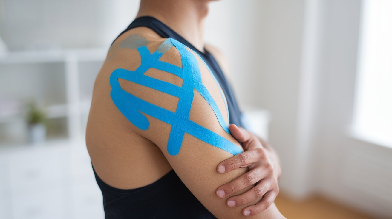
KT tape for shoulder rotator cuff problems is a popular topic among athletes, gym users, physical-therapy patients, and people experiencing shoulder discomfort. Kinesiology tape, often called KT tape, is an elastic adhesive tape that can be placed over and around the shoulder to provide sensory input and temporary support during movement. It is frequently used as an adjunct to rehabilitation, rather than as a stand-alone treatment for a rotator cuff injury.
The evidence available in 2026 presents a balanced picture. A 2026 systematic review and meta-analysis of 39 randomized controlled trials involving 2,481 participants found that kinesiology taping may provide small short-term improvements in shoulder function and pain, but the certainty of evidence was low to very low. The researchers concluded that KT tape may be considered alongside rehabilitation but should not replace education and exercise-based treatment.
What Is KT Tape for a Rotator Cuff Problem?
Kinesiology tape is designed to stretch and move with the body. Unlike rigid athletic tape, it does not completely restrict shoulder movement. When applied correctly, the tape can provide a light external stimulus to the skin while allowing the shoulder to move through its normal range.
The rotator cuff consists of four muscles and their tendons that help stabilize and move the shoulder joint. Problems involving these structures can include rotator cuff-related shoulder pain, tendinopathy, irritation, or injury. Symptoms may include pain when lifting the arm, discomfort when reaching overhead, weakness, or pain when sleeping on the affected side.
KT tape does not physically repair a torn tendon. Instead, people use it as a supportive technique that may make certain movements feel more comfortable during rehabilitation or daily activities. Earlier research has produced mixed results, and a systematic review specifically examining rotator cuff tendinopathy found insufficient evidence to establish KT as an effective stand-alone treatment.
How KT Tape May Help Shoulder Rotator Cuff Symptoms
The proposed benefits of KT tape for shoulder pain are primarily related to sensory feedback and movement rather than structural healing. The tape’s contact with the skin may alter how movement and discomfort are perceived, potentially making certain activities feel easier.
Research published in 2025 found statistically significant improvements in pain, shoulder flexion and abduction range of motion, and upper-limb function among participants with rotator cuff injuries who received kinesiology taping. However, the researchers also noted potential risk factors and advised interpreting the findings cautiously.
The newer 2026 systematic review provides useful context because it included substantially more randomized trials. Its conclusion was more conservative: KT produced, at best, small short-term benefits for shoulder function and pain during activity and at night, while effects on range of motion remained uncertain.
This means KT tape may be useful for some people as part of a broader rehabilitation plan, but it should not be viewed as a treatment that automatically heals a rotator cuff injury.
How to Apply KT Tape to the Shoulder
A common approach involves applying one or more strips around the shoulder while positioning the arm in a way that allows the tape to be placed without excessive tension. Exact taping techniques vary according to the symptoms, target area, and clinician’s preferred method.
Before applying KT tape, the skin should be clean, dry, and free of oils or lotions. The tape should be measured and rounded at the corners to reduce premature peeling. It is generally better to apply the tape smoothly rather than pulling aggressively on it.
For shoulder rehabilitation, a physical therapist may use a strip running along the shoulder or upper arm and additional strips designed to provide sensory input around the shoulder blade or deltoid region. Tape tension matters, and different techniques use different amounts of stretch. Because improper application can irritate the skin or fail to provide the intended effect, first-time users can benefit from having a qualified physical therapist demonstrate the technique.
KT tape should never be applied over open wounds, infected skin, significant rashes, or irritated areas. People with known adhesive allergies or very sensitive skin should also exercise caution.
KT Tape vs. Exercise for Rotator Cuff Recovery
One of the most important points when discussing KT tape and rotator cuff rehabilitation is that taping should not replace active treatment. Rotator cuff-related shoulder pain is commonly managed through progressive exercise, education, activity modification, and restoration of strength and movement.
A randomized clinical trial involving people with rotator cuff-related shoulder pain compared rehabilitation with and without KT tape. Both groups improved, but adding KT did not produce superior mid- or long-term results compared with an exercise-based rehabilitation program alone.
Research published in 2026 also supports the importance of exercise-based rehabilitation. A systematic review and meta-analysis comparing exercise therapy with passive conservative treatments examined randomized controlled trials involving rotator cuff-related shoulder pain.
Therefore, KT tape can be considered a complementary tool, particularly if it helps someone participate more comfortably in prescribed exercises or daily movement. The central rehabilitation program should remain focused on appropriate strengthening, mobility, neuromuscular control, and gradual return to activity.
How Long Can You Wear KT Tape?
The appropriate wearing time depends on the particular tape, application, skin sensitivity, activity level, and instructions from the manufacturer or healthcare professional. Many kinesiology-taping protocols leave tape in place for several days, but longer wear is not automatically better.
The skin should be checked regularly for redness, itching, burning, blistering, or irritation. If these symptoms develop, the tape should be removed carefully rather than continuing to wear it.
Removal should be gradual. Pulling the tape rapidly can irritate the skin, particularly when the adhesive has been worn for an extended period. Warm water or an appropriate adhesive-removal product can sometimes make removal more comfortable.
People should also remember that less pain while wearing tape does not necessarily mean the underlying injury has healed. Pain can change while tissue recovery is still occurring, so activity should remain consistent with professional rehabilitation guidance.
Safety, Limitations, and When to Seek Professional Help
KT tape is generally used as a low-risk supportive technique, but it is not appropriate for every person. Skin reactions, adhesive sensitivity, and incorrect application are among the main concerns. The tape should not be used as a substitute for diagnosis when shoulder symptoms are persistent, severe, or associated with significant weakness.
Medical assessment is especially important after a substantial injury, such as a fall or sudden force, or when there is an inability to raise the arm normally. Persistent night pain, progressive weakness, substantial loss of movement, swelling, deformity, numbness, or symptoms that do not improve should also be evaluated by a healthcare professional.
A rotator cuff problem can have several causes, and not every shoulder pain pattern is a rotator cuff injury. A clinician or physical therapist can help determine whether the symptoms are consistent with rotator cuff-related pain and whether imaging or additional treatment is appropriate.
The research also explains why KT tape should not be marketed as a guaranteed solution. A 2020 systematic review found no firm evidence that kinesiology taping was superior to sham taping, exercise, or passive treatments for shoulder pain and function.
Frequently Asked Questions About KT Tape and the Rotator Cuff
Does KT tape heal a rotator cuff tear?
No. KT tape does not repair a torn rotator cuff tendon. It may provide temporary sensory support or symptom relief but should not be considered a method for repairing damaged tissue.
Can KT tape reduce rotator cuff pain?
It may provide short-term pain relief for some people, but research findings are mixed. The 2026 evidence review found only small short-term benefits overall and rated the certainty of evidence as low to very low.
Can I use KT tape instead of physical therapy?
KT tape should generally be considered an adjunct rather than a replacement for rehabilitation. Exercise-based treatment remains an important component of recovery.
How should KT tape be placed for shoulder pain?
Application depends on the individual’s symptoms and the specific taping technique. A physical therapist or qualified healthcare professional can demonstrate the appropriate placement and tension.
Can I sleep with KT tape on my shoulder?
Some taping protocols leave the tape in place overnight, but skin tolerance varies. Check the skin regularly and remove the tape if irritation develops.
Is KT tape useful for rotator cuff tendinopathy?
It may provide a temporary supportive effect, but evidence does not establish it as a stand-alone treatment. Exercise-based rehabilitation remains central to management.
What is the main takeaway about KT tape for a shoulder rotator cuff problem?
KT tape can be a useful supportive rehabilitation tool for some people, particularly when it makes movement more comfortable. However, it should not be expected to repair a tendon or replace an appropriate exercise and rehabilitation program. The latest 2026 research supports this cautious, complementary role.

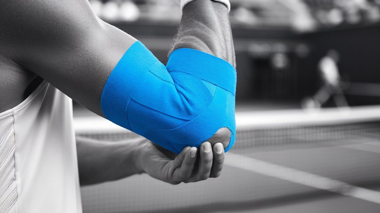
Tennis elbow is one of the most common overuse injuries affecting athletes, office workers, fitness enthusiasts, and individuals who perform repetitive arm movements. Whether the discomfort comes from tennis, weightlifting, typing, painting, or manual labor, the condition can significantly impact daily activities. One of the most popular non-invasive solutions used today is KT Tape for tennis elbow.
By providing targeted support to muscles and tendons without restricting movement, KT Tape has become a trusted recovery tool for people seeking relief from elbow pain. When applied correctly, it can help reduce strain, improve circulation, and support the healing process while allowing continued activity.
What Is Tennis Elbow and Why Does It Occur?
Tennis elbow, medically known as lateral epicondylitis, is a condition caused by repeated stress on the tendons that attach to the outer part of the elbow. Despite its name, it affects far more than tennis players.
The condition develops when the forearm muscles and tendons become overloaded through repetitive motions. Common causes include:
- Racquet sports
- Weight training
- Typing and computer work
- Painting
- Plumbing and construction work
- Gardening
- Repetitive lifting tasks
Typical symptoms include:
- Pain on the outer side of the elbow
- Weak grip strength
- Difficulty lifting objects
- Discomfort when shaking hands
- Pain while turning door handles
- Tenderness around the elbow joint
Without proper management, symptoms can persist for months and interfere with both work and recreational activities.
How KT Tape Helps Tennis Elbow Recovery
KT Tape, also known as kinesiology tape, is an elastic therapeutic tape designed to support muscles and joints while maintaining a full range of motion.
Unlike rigid athletic tape, KT Tape stretches and moves naturally with the body. When applied to the forearm and elbow area, it can provide several benefits.
Reduced Tendon Stress
The tape helps distribute mechanical load away from the irritated tendon. This reduces pressure on the damaged tissues and allows them to function more comfortably during movement.
Improved Circulation
The elastic properties of KT Tape gently lift the skin, creating additional space between tissues. This may encourage better blood flow and lymphatic drainage around the injured area.
Enhanced Muscle Support
KT Tape provides sensory feedback to the muscles, helping maintain proper movement patterns and reducing excessive strain.
Pain Reduction
Many users report decreased pain levels during activity because the tape supports the affected structures and may influence pain receptors in the skin.
Proper KT Tape Application for Tennis Elbow
Correct application is essential for achieving optimal results. While various taping methods exist, a common approach involves supporting both the forearm muscles and the affected tendon.
Step 1: Prepare the Skin
Before applying KT Tape:
- Clean the skin thoroughly
- Remove oils or lotions
- Trim excess arm hair if necessary
- Ensure the skin is completely dry
Step 2: Position the Arm
Extend the affected arm in front of the body with the palm facing downward. Gently bend the wrist toward the floor to stretch the forearm muscles.
Step 3: Apply the Main Strip
Place one end of the tape near the wrist without stretch. Apply the tape along the forearm toward the elbow using moderate tension. Finish near the outer elbow with no stretch on the final section.
Step 4: Add a Support Strip
Apply a shorter strip across the painful area on the outer elbow. This strip provides focused support where the tendon irritation is most prominent.
Step 5: Activate the Adhesive
Rub the tape firmly after application. The friction-generated heat activates the adhesive and improves tape longevity.
Most KT Tape applications can remain effective for several days, even during showering and exercise.
Benefits of Using KT Tape for Tennis Elbow
Many individuals choose KT Tape because it offers support without limiting mobility.
Maintains Natural Movement
Unlike braces or rigid wraps, KT Tape allows the elbow and wrist to move naturally. This makes it suitable for sports, exercise, and daily activities.
Lightweight and Comfortable
The tape is thin, breathable, and designed for extended wear. Most users quickly forget they are wearing it.
Supports Active Recovery
Instead of immobilizing the arm, KT Tape promotes movement while providing additional support to stressed tissues.
Versatile Use
It can be worn during:
- Tennis matches
- Golf rounds
- Gym workouts
- Office work
- Household chores
- Rehabilitation exercises
Cost-Effective Solution
Compared with repeated therapy sessions or specialized support devices, KT Tape offers an affordable option for ongoing management.
Best Exercises to Combine with KT Tape
While KT Tape can provide support, combining it with targeted exercises often produces better outcomes.
Wrist Extensor Stretch
Extend the arm with the palm facing down. Use the opposite hand to gently bend the wrist downward until a stretch is felt in the forearm.
Forearm Strengthening
Light resistance exercises help rebuild tendon capacity. Start with manageable weights and gradually increase intensity.
Grip Training
Using a soft stress ball or grip trainer can improve forearm strength and restore function.
Eccentric Wrist Extensions
These exercises focus on controlled lowering movements, which are commonly recommended for tendon rehabilitation.
Shoulder Stability Work
Improving shoulder strength can reduce excessive stress transferred to the elbow during activity.
Consistent exercise combined with KT Tape support can contribute to long-term improvement.
Common Mistakes When Using KT Tape for Tennis Elbow
Many people fail to achieve optimal results because of incorrect application techniques.
Applying Too Much Stretch
Excessive tension can cause skin irritation and reduce comfort. Moderate stretch is generally sufficient.
Ignoring Skin Preparation
Sweat, oils, and lotions can reduce adhesion and cause premature peeling.
Using Tape as the Only Treatment
KT Tape is most effective when combined with rest, strengthening exercises, and activity modification when necessary.
Applying Over Damaged Skin
Never place tape over cuts, rashes, infections, or irritated areas.
Continuing Painful Activities Without Modification
Although KT Tape may reduce discomfort, persistent overloading can delay recovery.
Who Can Benefit Most from KT Tape Tennis Elbow Support?
KT Tape is widely used by individuals experiencing mild to moderate tennis elbow symptoms.
Ideal candidates include:
- Tennis players
- Golfers
- Baseball athletes
- Weightlifters
- Office workers
- Musicians
- Tradespeople
- Fitness enthusiasts
- Manual laborers
Individuals recovering from repetitive strain injuries often find KT Tape useful during rehabilitation and return-to-activity phases.
However, severe pain, significant weakness, numbness, or symptoms lasting several months should be evaluated by a healthcare professional.
Choosing the Right KT Tape for Tennis Elbow
Several KT Tape options are available in 2026. Selecting the right version depends on activity level and personal preference.
Original Cotton KT Tape
Suitable for everyday wear and moderate physical activity.
Synthetic Performance KT Tape
Designed for intense workouts, swimming, and high-sweat conditions. It generally offers longer durability.
Pre-Cut Strips
Ideal for beginners who want simple application without cutting tape manually.
Professional Rolls
Provide flexibility for customized taping patterns and advanced support techniques.
Regardless of the version selected, proper application remains the most important factor in achieving effective support.
Conclusion
KT Tape for tennis elbow has become one of the most widely used support methods for managing elbow pain caused by repetitive strain. Its ability to provide targeted support while preserving natural movement makes it an attractive option for athletes and non-athletes alike. When combined with strengthening exercises, stretching routines, and smart activity management, KT Tape can help reduce discomfort, improve function, and support recovery. Consistent use, proper application, and a comprehensive rehabilitation approach often produce the best long-term results for individuals dealing with tennis elbow.
Frequently Asked Questions (FAQ)
Does KT Tape really help tennis elbow?
Yes. Many users experience reduced pain and improved support during activity when KT Tape is applied correctly.
How long can KT Tape stay on for tennis elbow?
Most KT Tape applications remain effective for three to five days, depending on activity level and skin conditions.
Can I exercise while wearing KT Tape?
Yes. KT Tape is specifically designed to support movement during sports, workouts, and daily activities.
Is KT Tape better than an elbow brace?
Both options can be effective. KT Tape offers more flexibility and mobility, while braces may provide stronger compression.
Can KT Tape cure tennis elbow?
KT Tape does not cure the condition by itself. It supports recovery and symptom management when combined with appropriate rehabilitation strategies.
When should I seek medical attention?
Consult a healthcare professional if symptoms become severe, persist for several months, or are accompanied by numbness, significant weakness, or loss of function.

Golden State Warriors vs Utah Jazz Match Player Stats 2026: Complete Box Score and Game Analysis

Voddler.co.uk: Complete Guide to the Website, Streaming History, Content, and Safety in 2026

SosoActive: Meaning, History, Websites, and What It Offers in 2026

Samantha Struthers Rader: Biography, Career, Family, and the Inspiring Journey of Dr. Sam

Stacie Zabka: Everything You Need to Know About William Zabka’s Wife

Imogen Lucie Age: Biography, Career, Height, Family, Net Worth, and Social Media (2026 Guide)
 News4 months ago
Samantha Struthers Rader: Biography, Career, Family, and the Inspiring Journey of Dr. Sam
- celebrity4 months ago
Stacie Zabka: Everything You Need to Know About William Zabka’s Wife
- celebrity3 months ago
Imogen Lucie Age: Biography, Career, Height, Family, Net Worth, and Social Media (2026 Guide)
- celebrity3 months ago
Ryan Kiera Armstrong: Biography, Movies, TV Shows, Career, Family, and Latest Updates (2026 Guide)
- celebrity3 months ago
Brecken Merrill Parents: Everything About His Family Background, Childhood, and Support System
- celebrity3 months ago
Priscilla Love Vanwinkle: Biography, Background, Family Connections, and Personal Story
- celebrity3 months ago
Moses Hacmon Net Worth: Biography, Career, Income Sources, and Financial Overview in 2026
- Blog3 months ago
@RedandWhiteMagz.com: A Complete Guide to the Digital Magazine and Blog in 2026